
Dott. Giannantonio Spena
Director of Neurosurgery Unit
Fondazione IRCCS Policlinico San Matteo
Pavia, Italy
Fondazione IRCCS Policlinico San Matteo
Pad. Nuovo Ospedale "DEA"
Piano +7 - Corpo A
Secretary + 39 382 502780
For Private Outpatient Appointment
Policlinico San Matteo: you can book an appointment by calling +39 382 501788 or by filling out the online form.
- Poliambulatorio Akesis viale Libertà 4, Pavia
+39 382 302996
PATHOLOGIES TREATED

INTRACRANIAL AND SPINAL VASCULAR PATHOLOGY
Vascular pathologies of the central nervous system are numerous. Those that most concern neurosurgeons are: cerebral aneurysms, arteriovenous malformations (AVMs), dural arteriovenous fistulas (DAVFs), and cavernomas.
All these pathologies can be discovered either by chance (during a diagnostic test performed for other reasons) or because they cause neurological disorders.
They can sometimes cause serious brain hemorrhages that require urgent treatment.
Cerebral aneurysms
Some examples of cerebral aneurysms operated on using the "clipping" technique, which consists of placing small tweezers at the base of the aneurysm sac, permanently closing it.


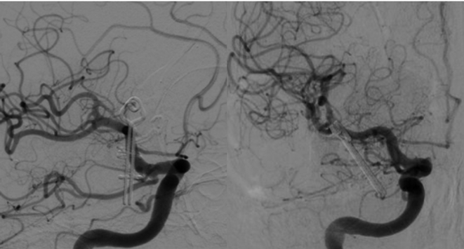
On the left is a photograph of an angiography of a young patient with multiple aneurysms (red arrows). On the right is the control angiography, which demonstrates the exclusion of the two aneurysms thanks to the clips that completely and permanently exclude them from blood entry.

Left: Angiography of a patient with an anterior communicating artery aneurysm (red arrow). Right: Postoperative CT angiography confirming the aneurysm was excluded using the clip.
Surgical exclusion (clipping) of right middle cerebral artery aneurysm
Typical surgical procedure involves clipping a right middle cerebral artery aneurysm. The aneurysm is located in a pocket between the frontal and temporal lobes. Therefore, much of the surgery involves reaching and preparing the aneurysm using microsurgical techniques. A small clip (6mm) is then placed at the base of the aneurysm, permanently preventing blood from entering.


Clipping of middle cerebral artery aneurysm (broad neck)
minipterional craniotomy

Giant cerebral aneurysms
Aneurysms can sometimes reach enormous dimensions. In these cases, in addition to posing the risk of rupture, they can behave like actual masses. Treatment therefore involves not only excluding the aneurysm from the arterial circulation but also removing the bulky sac. The video below shows a case of a giant aneurysm of the anterior cerebral artery (azygos).

Giant right carotid-choroidal cerebral aneurysm
A 68-year-old female patient was transported to the emergency room for a generalized seizure. Examinations revealed a giant, mostly thrombosed aneurysm originating from the internal carotid artery at the level of the anterior choroidal artery. A carotid flow diverter was placed to reduce flow into the sac and minimize risk during surgery. The patient then underwent craniotomy (with neurophysiological monitoring) and removal of the sac, as well as clipping of the neck. The choroidal artery was completely intact. Two years after surgery, the patient remains in perfect condition and has never had any further seizures.


Giant cerebral aneurysm of the posterior communicating artery


PICA (posterior inferior cerebellar artery) aneurysm - Vertebral.
The video below reports the case of a patient with an aneurysm originating from the left vertebral artery at the starting point of the PICA. Initially (three years earlier), the patient underwent endovascular surgery for the placement of a flow diverter. Unfortunately, the aneurysm was not occluded. Therefore, a craniotomy and aneurysm clipping were proposed.

Cerebral cavernomas
Cavernomas are vascular malformations consisting of tangles of dilated, thin vessels that resemble blackberries. Arterial blood flow does not enter these sacs at high pressure, thus differing from aneurysms and AVMs. However, cavernomas can still lead to cerebral hemorrhage or irritate the nervous system, causing seizures and, less frequently, neurological disorders.


The photo on the left shows an MRI and CT scan of a patient in critical condition due to a large hemorrhage in the third ventricle. The cause was a bleeding cavernoma. Surgery allowed evacuation of the hemorrhage and the cavernoma, as shown in the photo on the right.
The photo on the left shows an MRI of a very young patient with a large left temporal cavernoma that was causing numerous epileptic seizures. The young man underwent surgery to completely remove the cavernoma (images on the right), resulting in the disappearance of the seizures.


